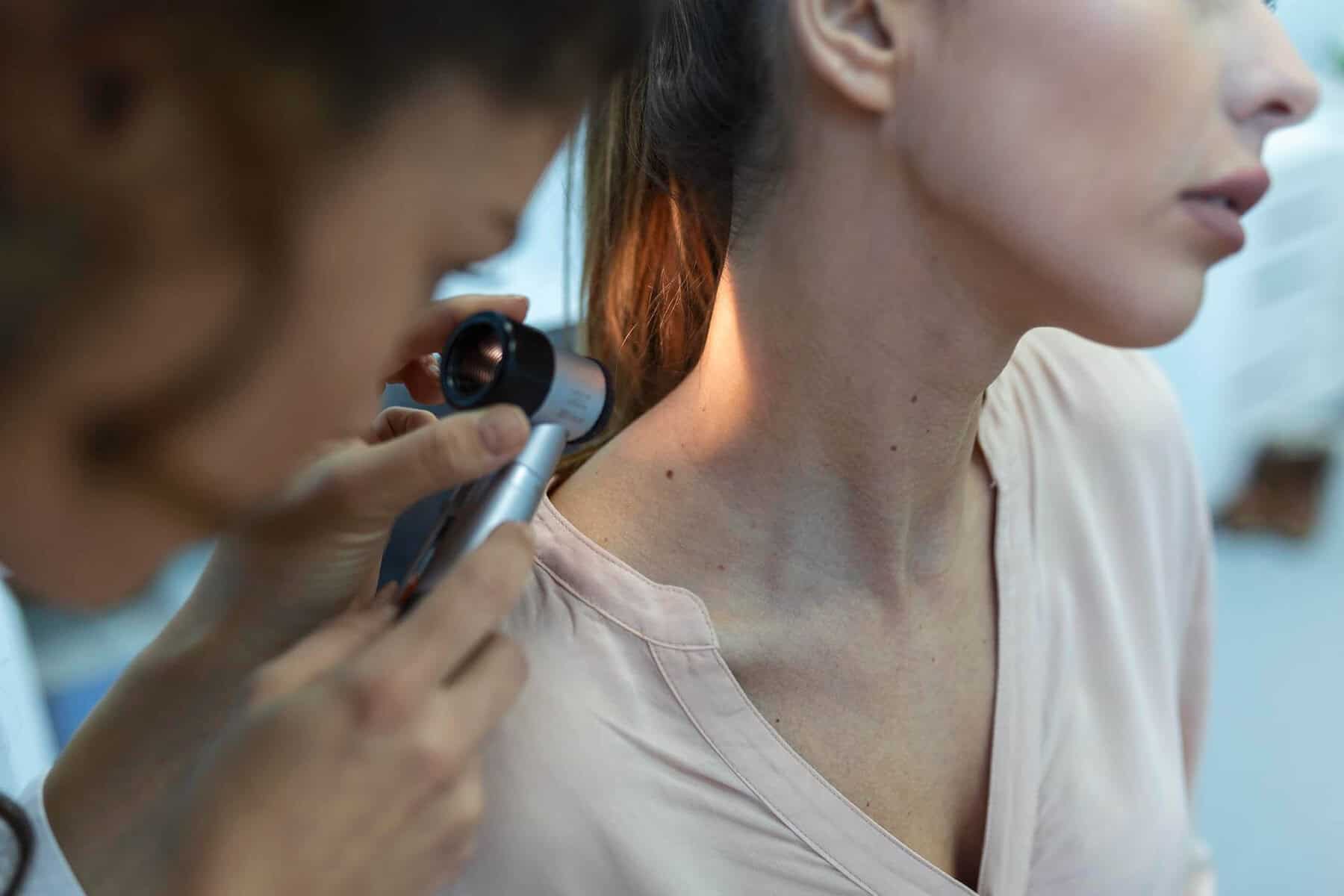
If you have recently had a mole removed, it is completely natural to feel anxious while waiting for the results. Many patients ask what happens to the tissue after it leaves the clinic, how long results take, and what different outcomes might mean.
A clear understanding of the mole removal results histology process can provide reassurance during the waiting period.
What Happens to a Mole After Removal?
Once a mole is removed during a mole removal procedure, it does not simply get discarded. If clinically indicated, the tissue is sent to a specialist laboratory for examination. This is known as histology.
Histology allows experts to look at the mole under a microscope to determine exactly what type of cells are present. It provides a definitive diagnosis, which cannot be confirmed by visual inspection alone.
Many patients ask, “What happens to a mole after removal?” The short answer is: it is carefully preserved, processed and analysed to ensure nothing important is missed.
The Histology Process: Step by Step
Understanding the histology pathway can make the waiting period feel more manageable.
1. Labelling and Preservation
Immediately after removal, the mole is placed in a preservative solution. It is carefully labelled with your details and sent securely to a specialist pathology laboratory.
2. Tissue Processing
In the laboratory, the sample is processed. This involves embedding it in wax, slicing it into extremely thin sections and placing those sections onto microscope slides.
3. Microscopic Examination
A consultant pathologist examines the slides under a microscope. They assess:
- Cell structure
- Cell arrangement
- Depth of the lesion
- Margins (whether the entire lesion has been removed)
- Any signs of abnormal or cancerous change
This detailed analysis produces your mole histology results.
How Long Does It Take to Get Mole Removal Results?
In most cases, results are available within 7–10 working days. Occasionally, complex cases may take slightly longer if additional tests are required.
You will be contacted once the report is reviewed. If results are urgent, you will be informed promptly. There is a clear and structured follow-up process for all abnormal findings.
What Does a Benign Result Mean?
Reassuringly, most mole removal results histology reports confirm that the lesion is benign.
A benign result means:
- The mole is non-cancerous
- No further treatment is required
- The removal was complete
- Routine skin monitoring is usually sufficient
For many patients, this confirmation brings significant peace of mind.
What Happens If Something Abnormal Is Found?
If histology shows abnormal or cancerous changes, the next steps depend on the type and extent of the finding.
Some lesions may be identified as early forms of skin cancers including basal cell, which are generally highly treatable when detected early.
In rarer cases, the report may identify features consistent with malignant melanomas. Early detection significantly improves outcomes.
If an abnormal result is identified:
- You will be contacted directly
- The findings will be explained clearly
- Further treatment, if required, will be arranged promptly
- A clear management plan will be outlined
It is important to emphasise that the majority of biopsied moles do not show melanoma.
Will I Be Contacted If My Results Are Urgent?
Yes. There is a structured follow-up pathway in place.
If your mole histology results require urgent attention, you will not be left waiting. The clinic will contact you directly to discuss next steps and arrange further care if needed.
If your result is benign, you will also be informed once the report has been reviewed.
Can I See My Own Histology Report?
Yes. Patients are entitled to view their results. If you would like a copy of your histology report for your records, this can be requested.
During your follow-up, the findings will be explained in clear, accessible language. Medical terminology in histology reports can be technical, so clarification is always available.
Aftercare While Waiting for Results
Waiting for results can be the most stressful part of the process. During this period:
- Follow your wound care instructions carefully
- Keep the area clean and dry
- Attend stitch removal if required
- Avoid excessive strain on the site
If you notice increasing redness, swelling or discharge at the biopsy site, contact the clinic for advice.
It is also helpful to remember that most mole removal results histology reports are benign.
Why Histology Matters
Histology provides certainty. While a dermatologist is highly trained in identifying suspicious features visually, microscopic examination confirms the diagnosis.
The process ensures that:
- Cancer is not missed
- Margins are clear where removal was intended to be complete
- Appropriate follow-up is arranged if necessary
It is a safeguard designed to prioritise patient safety.
Reassurance While You Wait
It is completely normal to feel anxious while waiting for mole removal histology results. However, most results confirm benign findings.
There is a clear process in place for reviewing, communicating and acting on results. You will not be left uncertain about what comes next.
If you have recently undergone mole removal and have questions about your results, or if you are considering assessment for a changing mole, book a consultation at The Forbury Clinic for expert advice and follow-up.