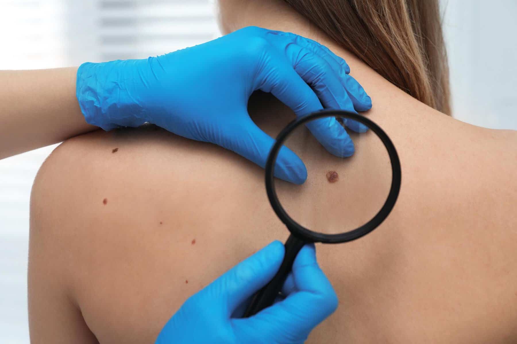
If you have been advised to consider mole removal, you may quickly encounter two terms: shave biopsy and excision biopsy. For many patients, the immediate question is not whether to remove the mole, but which method is right.
Understanding the difference between these mole biopsy types can help you feel confident about the decision. Here we explain the differences between shave biopsy vs excision biopsy and when each is used, so you can make an informed choice about your care.
Why This Decision Matters
When patients search for “mole removal biopsy,” they are usually weighing up two concerns:
Is the mole suspicious?
What will the scar look like afterwards?
Both shave biopsy and excision biopsy are safe and effective. However, they serve slightly different purposes. The method chosen depends on whether the mole appears benign or potentially malignant, its size, depth and location, and whether complete removal is required for diagnostic reasons.
If you are considering mole removal, understanding this distinction is essential.
What Is a Shave Biopsy?
A shave biopsy involves removing the raised portion of a mole using a fine surgical blade. The procedure is performed under local anaesthetic, and stitches are usually not required.
When Is a Shave Biopsy Used?
Shave biopsy is commonly used for:
- Raised, clearly benign moles
- Cosmetic removal of non-suspicious lesions
- Superficial skin growths
It is particularly suitable when the mole is elevated above the skin surface and there is no clinical concern for deeper invasion.
Advantages of Shave Biopsy
- Quick procedure
- No stitches in most cases
- Generally shorter healing time
- Often leaves a flat, small scar
Patients often ask, “Which biopsy method leaves less scarring?” For clearly benign, raised moles, shave biopsy can produce a cosmetically pleasing result because it avoids a deeper cut.
Does a Shave Biopsy Provide Enough Tissue for Histology?
In many benign cases, yes. However, when there is suspicion of melanoma or deeper pathology, a shave biopsy may not capture the full depth of the lesion. In those cases, excision biopsy is usually preferred.
What Is an Excision Biopsy?
An excision biopsy removes the entire mole, including a small margin of surrounding skin. This is a full-thickness removal that allows for complete histological analysis.
The area is numbed with local anaesthetic, the lesion is surgically removed, and stitches are placed to close the wound.
When Is an Excision Biopsy Used?
Excision biopsy is generally preferred for suspicious moles where deeper tissue sampling matters. It is typically recommended when:
- A mole has changed in size, shape or colour
- There are irregular borders or uneven pigmentation
- Melanoma is suspected
- Complete removal is required for diagnosis and treatment
If the mole raises concern, excision biopsy provides the most accurate histology as it preserves the full depth of the lesion.
Advantages of Excision Biopsy
- Removes the entire lesion
- Provides full-depth tissue for biopsy histology
- Reduces risk of incomplete sampling
- Often definitive treatment if benign or early malignant
Because excision biopsy involves stitches, healing may take slightly longer compared to a shave biopsy. However, in suspicious cases, diagnostic accuracy takes priority over cosmetic speed.
Shave Biopsy vs Excision Biopsy: Side-by-Side Comparison
| Feature | Shave Biopsy | Excision Biopsy |
|---|---|---|
| Depth of removal | Surface-level | Full thickness |
| Stitches required | Usually no | Yes |
| Healing time | 1–2 weeks | 1–2 weeks for stitches; scar matures over months |
| Histology accuracy | Suitable for benign lesions | Preferred for suspicious moles |
| Scarring | Often minimal and flat | Linear scar |
| Best for | Raised benign moles | Suspicious or changing moles |
When comparing shave biopsy vs excision biopsy, the most important difference is depth. Excision biopsy provides more comprehensive tissue for analysis.
Which Biopsy Method Is Used for Suspicious Moles?
If a mole appears clinically suspicious, excision biopsy is generally recommended. This is because melanoma assessment requires evaluation of depth, margins and structural features.
Shave biopsy may not always provide enough information in these cases. Therefore, where diagnostic certainty is critical, excision is typically the safer and more appropriate approach.
Patients often search, “Which biopsy method is used for suspicious moles?” In most cases, the answer is excision biopsy.
How Does a Dermatologist Decide Which Method to Use?
The decision is always based on careful clinical assessment.
Procedures are performed by consultant dermatologist Sacha Goolamali at The Forbury Clinic, considering:
- The ABCDE features of the mole
- Whether the lesion is flat or raised
- Dermoscopic findings
- Patient age and medical history
- Cosmetic location
- Clinical suspicion of malignancy
If the mole is clearly benign and raised, shave biopsy may be sufficient. If there is uncertainty or suspicion, excision biopsy is usually recommended.
Every decision balances diagnostic safety and cosmetic outcome.
Healing and Aftercare
Regardless of the mole biopsy type chosen, both procedures are performed under local anaesthetic and patients return home the same day.
After Shave Biopsy
- A small dressing is applied
- The site heals gradually over 1–2 weeks
- No stitch removal required
After Excision Biopsy
- Stitches remain in place for 7–14 days
- The scar may feel firm initially
- Scar maturation continues for several months
Most patients ask whether mole removal biopsy is painful. The procedure itself is not painful due to local anaesthetic. Mild tenderness afterwards is normal and manageable.
Does One Method Guarantee Better Cosmetic Results?
Not necessarily. Cosmetic outcome depends on:
- Mole size
- Location on the body
- Skin type
- Healing tendency
Shave biopsy often leaves a flatter scar, but in some cases may result in a slight indentation. Excision biopsy leaves a fine linear scar, which fades over time.
Your dermatologist will discuss expected scarring before proceeding.
What Happens to the Mole After Removal?
Both shave biopsy and excision biopsy samples are sent for histological examination when clinically indicated. This laboratory analysis confirms the diagnosis.
Results typically take several working days. If malignancy is detected, further treatment planning is arranged promptly.
Making the Right Decision
If you are deciding between shave biopsy vs excision biopsy, the most important factor is clinical appropriateness. Cosmetic concerns matter, but diagnostic safety comes first.
For clearly benign, raised moles, shave biopsy can be efficient and cosmetically favourable. For suspicious moles, excision biopsy offers greater diagnostic confidence.
Book a Consultation
Whether you are considering cosmetic mole removal or concerned about a changing lesion, consultant-led evaluation ensures you receive the right procedure for your needs.
To discuss your options with a consultant dermatologist, contact The Forbury Clinic today.